Higher Than Normal Mitochondrial DNA Movement May Play Role in Colorectal Cancer


Patients with colon and rectal cancer have higher than normal pieces of mitochondrial DNA in their larger DNA portfolio, which is known as nuclear DNA, according to a study.
The University of Alabama, Birmingham study, “Migration of mitochondrial DNA in the nuclear genome of colorectal adenocarcinoma,” was published in the journal Genome Medicine.
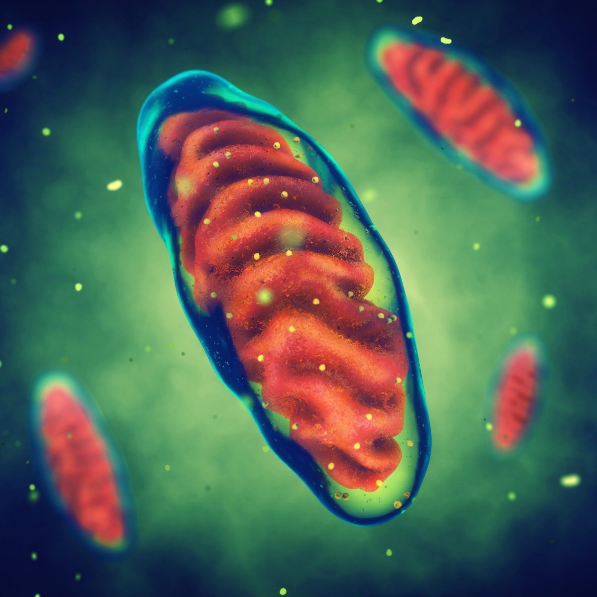
Our cells carry two genomes. One, packed in the cell nucleus, contains the bulk of our genomic information, with thousands of genes. The other, the mitochondria DNA, is located inside a small organelle, the mitochondria, and has a significantly lower number of genes — only 37.
Colorectal cancer displays the same genomic instability as all cancers: an increased tendency for the genome to have alterations, or mutations. But colorectal cancer also carries abnormalities in mitochondrial DNA.
Whether there was any interaction between the two genomes — nuclear and mitochondrial — was unknown until recently. Researchers at the University of Alabama at Birmingham have discovered higher than normal pieces of mitochondria DNA in the nuclear DNA of cells from colorectal cancer patients.
They analyzed genome information from patients with colon and rectum cancer who participated in the Cancer Genome Atlas. The goal was to determine the number and distribution in the genome of pieces of mitochondria DNA, or what scientists call mitochondria DNA insertions.
The colorectal cancer cells contained almost 4 1/2 times more pieces of mitochondrial DNA in the nuclear DNA than healthy cells, the researchers discovered.
Importantly, they found a correlation between the number of insertions and higher patient death rates. The number of insertions in colorectal tumors was higher in women than men.
The team concluded that a higher than normal level of insertions of mitochondrial DNA in the nuclear genome may play an important role in tumor development. But more research is needed to confirm their findings, they emphasized.
Previous studies involving the yeast Saccharomyces cerevisiae indicated that a gene called YME1 suppressed the migration of mitochondrial DNA to the nuclear genome.
The equivalent gene in humans is called YME1L1, and the Alabama esearchers decided to analyze it as well. They found that in colorectal tumors, the YME1L1 gene is frequently mutated, meaning that it has many errors in its sequence. The same pattern was detected in other cancers.
When the scientists deleted YME1L1 from human cells, they saw an increase in mitochondrial DNA insertions in the nuclear genome. The results indicate that YME1L1 is responsible for suppressing mitochondrial DNA transfer toward the nucleus. It is the first human gene that has been identified as playing such a role.
Overall, the findings suggest that higher than normal insertions of mitochondrial DNA in the nuclear genome may play a key role in cancer development. The insertions may also be a biomarker for cancer, the researchers said. Its use as a biomarker would require faster and more accurate ways to detect and analyze insertion of mitochondrial DNA into cell genomes, however, the team said.
With this in mind, the Alabama scientists and colleagues at the Department of Plant Pathology at Kansas State University decided to develop a new molecular tool to analyze the insertions faster and more efficiently. It uses fluorescent probes to mark specific mitochondria DNA. The study, “Single molecule mtDNA fiber FISH for analyzing numtogenesis,” was published in the journal Analytical Biochemistry.
“This novel technique should help distinguish and monitor cancer stages and progression, aid in elucidation of basic mechanisms underlying tumorigenesis, and facilitate analyses of processes related to early detection of cancer, screening and/or cancer risk assessment,” researchers wrote.