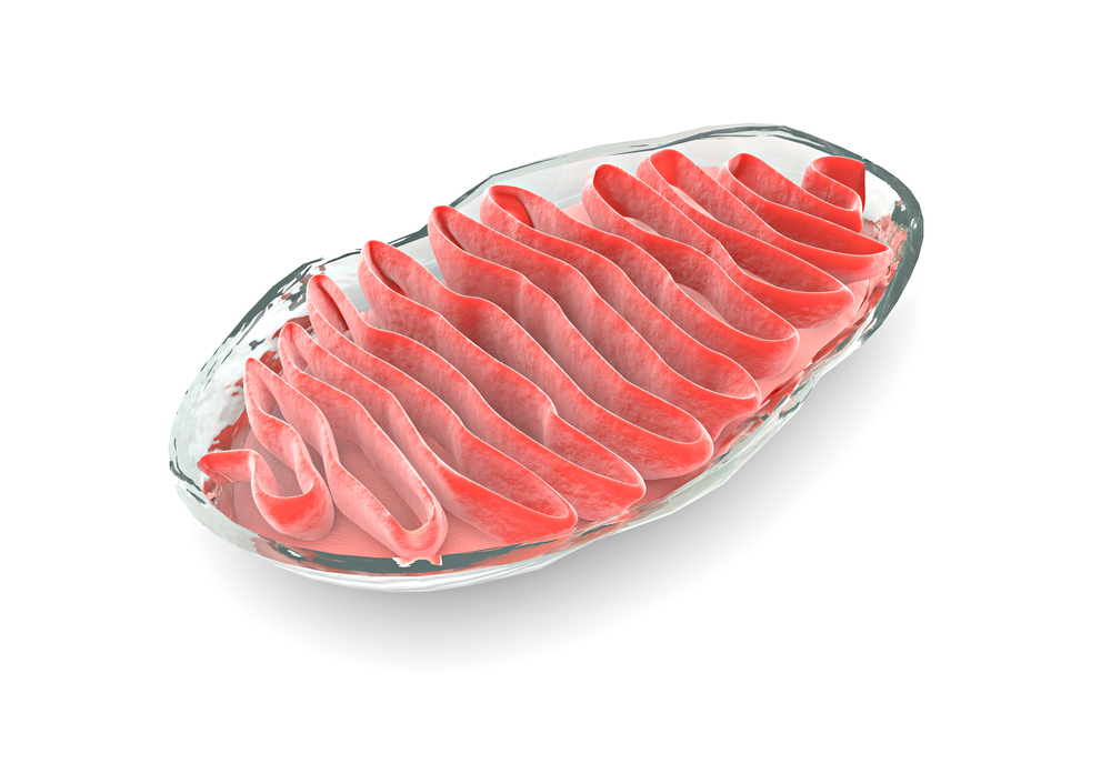
Effective Therapies and Diagnostics for Mitochondrial Disease Remain an Unmet Need

Mitochondrial myopathy is a collective term for diseases related to defective mitochondria in cells. In patients with mitochondrial myopathy, mitochondria are not able to undergo normal oxygen consumption and energy production due to mutations in mitochondrial DNA (mtDNA) or nuclear DNA that affect the mitochondrial machinery of cells. Diseases appear most often in children as young as infants, but according to the “Facts About Mitochondrial Myopathies” document, which was published by the Muscular Dystrophy Association, some can become apparent in adulthood as well. These diseases are relatively rare, which has unfortunately impacted the number of treatment options available for individuals affected by mitochondrial myopathy.
Most of the treatments available for mitochondrial disease are directed toward treating the symptoms of disease and not necessarily the disease itself. Heart problems, strokes, seizures, migraines, deafness, and diabetes all have long-standing, effective treatments (with these conditions being common complications of mitochondrial disease). However, since complication-specific treatment options address only the complications and not the disease itself, other effects of mitochondrial myopathy are not mitigated by these options.
Alternatively, dietary supplements that promote efficient use of functioning mitochondria are sometimes included in part of mitochondrial myopathy patients’ treatment regimens. These supplements focus on three molecules of energy production in cells: creatine, carnitine, and coenzyme Q10. The efficacy of administering these supplements to patients to increase the efficiency of mitochondria function is debated, as many supplements do not seem to produce a robust therapeutic effect.
As a result, there is still a significant unmet need for therapies that treat mitochondrial myopathy. On the cusp of new treatment options, there are currently 38 registered clinical trials that seek or have sought to administer interventional treatment to mitochondrial myopathy patients. The majority of treatments are in Phase 1 or Phase 2 clinical trials, which come before the Phase 3 clinical trials that can lead to market approval by the Food and Drug Administration (FDA).
One drug in Phase 1/2 clinical trials is MTP-131 (Bendavia) from Stealth BioTherapeutics Inc. The molecule is administered through intravenous injection and was developed to reduce oxidative stress in cells. Once Bendavia reaches the interior of a cell, it targets a lipid known as cardiolipin, which is found exclusively in the inner membrane of the mitochondria to ensure optimal function of enzymes involved in mitochondria energy production. During the preclinical development stage, a presentation at the American Heart Association Meeting described how Bendavia was able to improve mitochondria respiration and rate of energy production in dogs. The current clinical trial will investigate safe dosing of Bendavia in humans.
In Phase 2 clinical trials is another drug, RTA 408, from Reata Pharmaceuticals. The drug is being developed for mitochondrial myopathy. RTA 408 is thought to work through activating the Nrf2 cell signaling pathway to increase mitochondrial biogenesis, reduce harmful reactive oxygen species, and increase mitochondrial efficiency. These three contributions may improve patients’ overall energy metabolism and relieve disease symptoms. During the two-part MOTOR clinical trial, an estimated 56 mitochondrial myopathy patients will be randomized into different treatment groups of either RTA 408 capsules or placebo capsules. The first part of the study will investigate the safety of increasing doses of RTA 408, and the second part of the study will investigate the efficacy of given doses of RTA 408. Efficacy will be measured through changes in peak workload during exercise and changes in the distance walked during a six-minute walk test.
A drug in more advanced Phase 2/3 clinical trials is RP103 (cysteamine) from Raptor Pharmaceuticals. The molecule is involved in converting cystine into cysteine, which is a key component of glutathione (GSH) synthesis. GSH is the main antioxidant in cells, and the limiting reagent in its synthesis is available cysteine. By administering RP103 to patients with Leigh syndrome, it is predicted that aberrant reactive oxygen species generation will be mitigated to reduce the level of oxidative damage in cells. The Phase 2/3 trial (RP103-MITO-001) is an open-label, dose escalating study to assess the safety, tolerability, efficacy, pharmacokinetics, and pharmacodynamics of RP103, and a long-term extension component to assess the efficacy of RP103 over time.
In addition to potential prescription drug-based treatments for mitochondrial myopathy, researchers are looking at genetic and cell-based approaches to treatment. A large collaborative research effort conducted in the United States and United Kingdom recently found that cells taken from patients with mtDNA mutations can be genetically corrected. “Metabolic Rescue in Pluripotent Cells from Patients with mtDNA Disease,” which was published in the journal Nature, described the procedure used by the researchers to revitalize cells from patients with mitochondrial encephalomyopathy and stroke-like episodes (MELAS) and Leigh syndrome. After turning the cells into stem cells, the researchers corrected the genetic mutations and monitored oxygen consumption and energy production in the cells. Reprogramming the cells resulted in normal metabolic function regarding oxygen consumption and energy production. Yet despite these results, multiple future research studies are necessary before these approaches can be translated into clinical use.
Not only are mitochondrial myopathies in need of appropriate treatments to alleviate symptoms in patients, but they also are in need of optimized diagnostic techniques. According to “Techniques and Pitfalls in the Detection of Pathogenic Mitochondrial DNA Mutations,” which was published in The Journal of Molecular Diagnostics, mtDNA is responsible for coding 5% of the proteins found in the mitochondria. Any mutation on mtDNA, of which there are between two and ten copies within a single mitochondria, can cause mitochondrial myopathy if the mutation leads to aberrant production of proteins integral to the oxidative phosphorylation system that creates energy in cells.
RELATED: Mitochondria and the Many Disorders That Compose “Mitochondrial Disease”
Detecting mutations in mtDNA has been recognized as an important step in diagnosing individuals with mitochondrial myopathy. However, some of the different molecular biological techniques that are used to detect mutations can produce unclear or easily misinterpreted results. Minimizing the error in interpreting results and developing techniques that produce as little overlap in differentiating one myopathy from another are thus essential to help patients with mitochondrial myopathy.
Deletions, duplications, and rearrangements are common mutations in mtDNA. Interestingly, the number of base pairs deleted or the location on a mtDNA gene have no influence on specific disease phenotypes. To detect deletions, Southern blot analysis is most common, where mtDNA is extracted and separated into bands on a gel based on size. If normal and mutated mtDNA are compared side-to-side, the mutated mtDNA sample will show bands or “smear” patterns that are not present in the normal sample. Unfortunately, if the sample of mutated mtDNA extracted from cells is not large enough (cells commonly have a mix of both mutated and normal mtDNA), Southern blot analysis is less effective and additional modes of detection are necessary.
Toward the goal of understanding the differences among mitochondrial myopathies based on the different mutations present, animal models of disease have become useful means to study individual diseases. “A wide range of strategies have been developed and utilized in attempts to mimic human mtDNA mutation in animal models,” wrote the authors of “Animal Models of Human Mitochondrial DNA Mutations,” which was published in the journal Biochimica et Biophysica Acta. “Use of these animals in research studies has shed light on mechanisms of pathogenesis in mitochondrial disorders, yet methods for engineering specific mtDNA sequences are still in development.”
As an added bonus of developing animal models that mimic human mitochondrial myopathy, animal models can be used to test potential treatments during preclinical development. As more pharmaceutical and biotechnology companies recognize the large unmet need in mitochondrial myopathy treatment, they will likely use these animal models of disease to test their compounds before initiating clinical trials in humans. If results of these research efforts are positive and show signs of positive benefits in patients with mitochondrial myopathy, the drugs may be approved by the FDA for marketing, and the unmet need will slowly be addressed.


