University of Arkansas Team Awarded NIH Grant to Find Better Diagnostic Tools for Mitochondrial Diseases


An interdisciplinary University of Arkansas research team has been awarded a grant by the National Institutes of Health to find better and faster diagnostic tools for mitochondrial diseases.
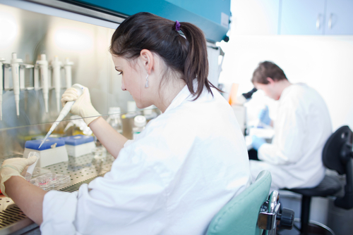
The team — led by Kyle Quinn, assistant professor of biomedical engineering, and Shilpa Iyer, assistant professor of biological sciences — will use the two-year grant of $395,112 to find more sensitive ways to detect problems in mitochondria. Early disease diagnosis can improve chances for intervention, and provide opportunities for patient enrollment in clinical trials. The diseases primarily affect children.
Diagnosis is often difficult due to the complex nature of mitochondrial diseases. Patients, on average, see more than eight physicians, undergo multiple tests, and receive misdiagnoses before being correctly diagnosed, according to a 2018 study.
Mitochondrial diseases arise from malfunctions in the mitochondria, the cell’s powerhouse that specializes in energy production. Most commonly affected systems are those that require higher amounts of energy, such as the nervous, cardiovascular, and gastrointestinal systems.
Such disorders are difficult to diagnose because patients often simultaneously have other chronic conditions — diabetes, heart disease, and neurodegenerative conditions, for example — that are common in the general population. Because cells can carry a mix of working and non-working mitochondria, defects can be masked by the presence of the functional mitochondria.
The grant will enable the scientists to assess a patient’s cell metabolism using label-free multiphoton imaging techniques being developed in Quinn’s lab.
Instead of injecting dyes or radiotracers, the researchers will use the natural fluorescence of mitochondria to visualize its function. The aim is to assess how the non-invasive optical method can complement current diagnostic approaches, which often necessitate painful biopsies of a patient’s muscle.
Iyer and her research group have done extensive work in the mitochondria field, including creating disease-specific stem cells derived from a patient’s own skin cells. Overall, the group is focused on understanding the role of bioenergetics and mitochondrial genetics in normal and diseased skin, nerve, and muscle cells — critical cell types affected in mitochondrial diseases.
Such investigations led Iyer to Quinn’s team, which has been studying skin wound healing through advanced optical imaging.
“It is a natural fit to combine our imaging work on skin metabolism with Dr. Iyer’s expertise in mitochondrial diseases and bioenergetics,” Quinn said in a press release.
Both scientists are motivated by a desire to improve the lives of patients with mitochondrial disease.
“It’s one of the most meaningful things we can do for children who suffer from such diseases,” said Iyer.