New Method Measures Mitochondrial Capacity to Produce Energy in Individual Muscles


A noninvasive method for analyzing the effectiveness of mitochondrial energy production in individual muscle groups shows promise to improve assessments in people with mitochondrial disease.
Researchers hope the new method will complement current methods, based on magnetic resonance spectroscopy, to improve diagnostics, long-term monitoring, and evaluation of treatment response in individuals with different types of mitochondrial diseases.
The technique was described in the study, “Muscle oxidative phosphorylation quantitation using creatine chemical exchange saturation transfer (CrCEST) MRI in mitochondrial disorders,” recently published in the journal JCI Insight.
Although oxidative phosphorylation capacity — the ability of mitochondria to produce energy — can vary widely between different muscle groups, current methods for assessing mitochondrial output have not been able to distinguish well between different muscles.
So researchers developed a new method called creatine chemical exchange saturation transfer, or CrCEST, that can assess metabolism in living organisms. A slower decline in CrCEST after exercise reflects a lower capacity for oxidative phosphorylation — in other words, a lower mitochondrial energy-making ability.
Now, scientists at the University of Pennsylvania evaluated the technique for measuring the decline in muscle free creatine after exercise in people with genetically determined mitochondrial diseases.
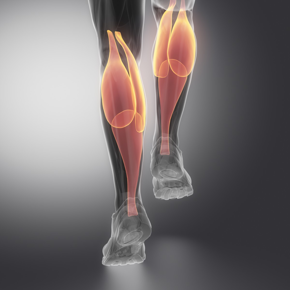
Researchers recruited 13 patients with mitochondrial disease and 14 healthy controls, and measured CrCEST both at rest and following exercise in three different calf muscles. Participants were of similar age and weight, and both groups were composed of mainly Caucasian people, of which about 60 percent were women.
The study found there was no difference between patients and controls in the crCEST levels at rest.
After exercise, free creatine, measured by CrCEST, increased in both groups. The amount of increase differed by muscle type, but was higher in patients with mitochondrial disease. The difference was not statistically significant, however.
The research team found that it took the different muscles between 1.4 and 1.7 minutes to get back to pre-exercise levels of free creatine in control individuals. For people with mitochondrial disease, the range was 2.0 to 2.2 minutes — a significant difference for one of the muscles.
The team also found that people who regularly exercised had a shorter time to recovery of free creatine, suggesting that the mitochondrial energy-making capacity gets better with exercise; this is also true in mitochondrial disease patients.
“Our work lays a rational foundation to develop CrCEST as a potential noninvasive imaging-based biomarker of OXPHOS capacity that may be useful in the diagnosis, long-term monitoring, and/or evaluation of response to clinical treatments in individuals with diverse types of mitochondrial diseases,” the authors concluded.