Scientists May Have Found New Mechanism Behind Mitochondrial Disease Development


Errors in the chemical processing that takes place in the production of mitochondrial proteins may play a role in the development of certain mitochondrial diseases, scientists from Japan have found.
The study, “Defective Mitochondrial tRNA Taurine Modification Activates Global Proteostress and Leads to Mitochondrial Disease,” was published in the journal Cell Reports.
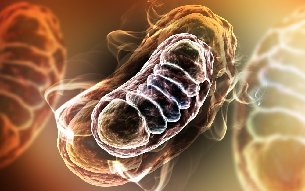
Human cells have two different types of DNA — mitochondrial and nuclear. Mitochondrial DNA has the necessary genetic information required to produce 22 different mitochondrial transfer RNAs (mt-tRNAs), which are part of the machinery responsible for making mitochondrial proteins, a process called mitochondrial translation. These proteins go on to play important roles in mitochondrial function.
If someone is born with mutations in their mitochondrial DNA that affect the production of mt-tRNAs, it can have an impact on a variety of mitochondrial proteins. This kind of mutation can lead to severe mitochondrial dysfunction and multisystemic failures in the body.
Mitochondrial tRNAs go through various chemical modifications which play an important part in their proper functionining. To date, 15 types of modifications have been identified at 118 positions in the 22 mt-tRNAs.
A subset of these mt-tRNAs contain a unique modification derived from taurine, a non-essential amino acid found in most animals. Taurine modification has been shown to play a role in stabilization of mitochondrial translation.
Mutations in the genes MTO1 and GTPBP3, which are known to play a role in taurine modification in yeast, are associated with severe mitochondrial dysfunction in human patients.
Therefore, researchers sought to determine the exact role of taurine modification in mitochondrial dysfunction using cellular and mouse models that lacked the Mto1 protein.
Researchers first showed that Mto1 was indeed responsible for taurine modification in mammals, which had previously not been proven.
Next, they showed that a deficiency of the protein Mto1 severely impaired the ability of the mitochondria to produce proteins, as well as to carry out respiration — the process by which cells get their energy.
Interestingly, researchers reported that in the Mto1-deficient cells, one of the structural components of the mitochondria — the inner mitochondrial membrane — had collapsed.
In addition, proteins that normally enter the mitochondria were no longer able to enter due to the collapsed membrane. This led to an accumulation of these mitochondrial proteins in the rest of the cell, which led to cell toxicity.
Researchers believe this may be the mechanism behind cell toxicity in patients with mitochondrial myopathy, encephalomyopathy, lactic acidosis, stroke-like symptoms (MELAS), or myoclonic epilepsy with ragged red fibers (MERRF).
This toxicity was rescued by using a compound called TUDCA (ursodeoxycholic acid), which led to a significant improvement in functional mitochondrial proteins in Mto1-deficient cells and mouse models.
“TUDCA, which has been shown to be effective in cell and [animal] models, is used as a remedy for liver diseases in Italy and other European countries, so its pharmaceutical safety has already been confirmed,” Prof. Wei Fan-Yan of Kumamoto University, leader of the research project, said in a press release.
“We are planning to conduct clinical trials in the near future to determine whether TUDCA is an effective remedy for mitochondrial diseases in humans,” he added.